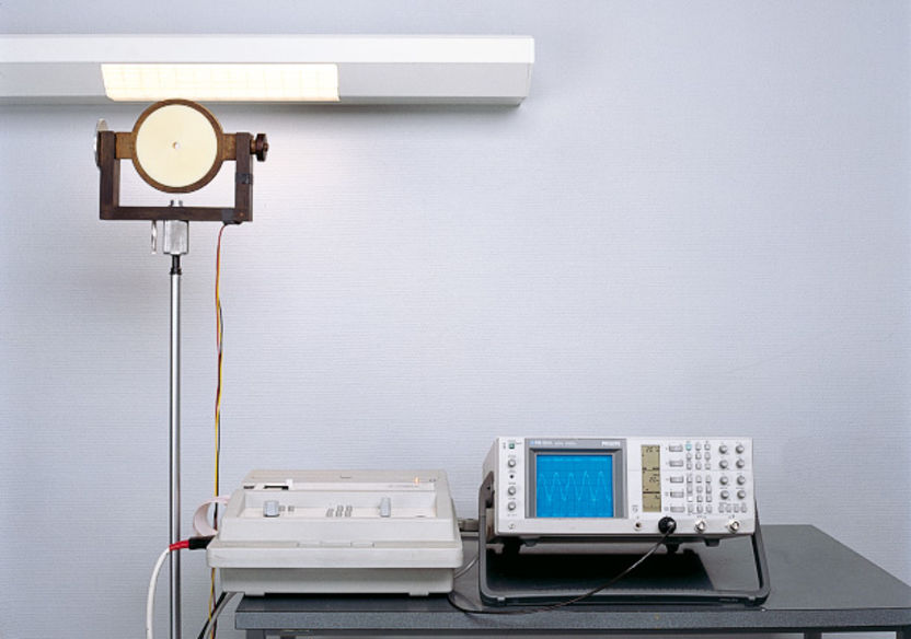
Figure 3.130: Electrocardiograph with standard exploring coil for measuring magnetic induction of a hospital room installation unit
Postbox 1960
D-59753 Arnsberg

Figure 3.130: Electrocardiograph with standard exploring coil for measuring magnetic induction of a hospital room installation unit
Live cells generate electric potentials due to their metabolic processes. Through analysis of their rhythm, frequency and form (amplitude), the physician gathers important information on brain function (EEG – electroencephalogram), heart function (ECG – electrocardiogram), muscle function (EMG – electromyogram) and other organ functions. In rooms where body activity potentials are recorded, interference caused by electric or magnetic fields must be avoided. This particularly applies to
EEG, ECG and EMG rooms,
intensive care examination and monitoring rooms,
heart catheter and operating rooms.
Electric fields are weakened by shielding measures like conductive wallpaper, grids (Faraday cage) etc. to an extent that mostly eliminates interference. Interference which can be caused by mains-frequency magnetic fields from e.g. by power lines, elevator engines and magnetic control gear in fluorescent luminaires can be effectively avoided by reducing the magnetic fields of interference sources and appropriate distances between measuring circuit and source (e.g. control gear).
Minimum distances are
ca. 75 cm for inductive control gear, and
ca. 30 cm for electronic control gear.
This way, the maximum induction values specified by DIN VDE 0100-710 "Low-voltage electrical installations – Part 7-710: Medical locations"
for ECG BSS = 4 · 10-7 Tesla = 0,4 μT
for EEG BSS = 2 · 10-7 Tesla = 0,2 μT
for EMG BSS = 1 · 10-7 Tesla = 0,1 μT
are not exceeded.
Figure shows a measuring method for easy on-site magnetic induction measurement.
EN ISO 11197 "Medical supply units" describes a block diagram for a corresponding measuring device (q.v. chapter "Medical-use rooms").